
In the realm of medical diagnostics, imaging techniques play a crucial role in accurate diagnosis and treatment planning. Among these, Dexa scans and MRIs stand out for their unique applications and benefits. This article delves into the specifics of each imaging modality, particularly focusing on their use in Prestons, and compares their respective roles in modern healthcare.
Understanding Dexa Scans: Precision in Bone Health
What is a Dexa Scan?
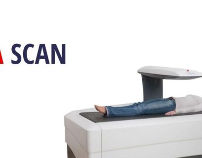
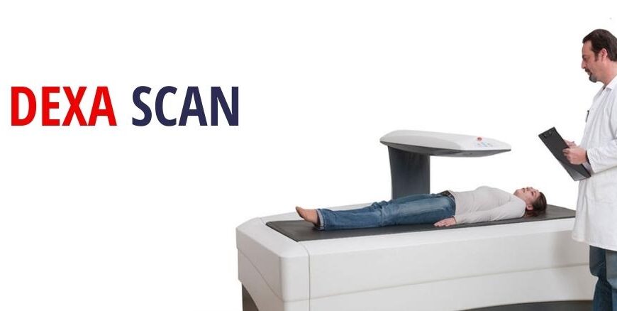
A Dual-Energy X-ray Absorptiometry (Dexa) scan is a specialized imaging technique used primarily to measure bone mineral density (BMD). Unlike standard X-rays, which are often used for detecting fractures or assessing general bone health, Dexa scans provide precise measurements of bone density. This is crucial for diagnosing osteoporosis and other bone-related conditions.
How Does a Dexa Scan Work?
During a Dexa scan, two X-ray beams at different energy levels pass through the bone. By comparing the amount of X-rays absorbed by the bone to the amount absorbed by soft tissue, the machine calculates the density of the bone. The scan is quick, non-invasive, and involves minimal radiation exposure, making it a safe choice for repeated assessments.
Applications of Dexa Scans
- Osteoporosis Diagnosis: Dexa scans are the gold standard for diagnosing osteoporosis, a condition characterized by weakened bones that are more susceptible to fractures.
- Fracture Risk Assessment: By measuring bone density, Dexa scans help predict the risk of fractures, especially in postmenopausal women and older adults.
- Body Composition Analysis: In addition to bone density, some advanced Dexa scanners can measure body fat and lean muscle mass, providing a comprehensive view of a patient’s health.
MRI in Prestons: Advanced Imaging for Comprehensive Diagnosis
What is an MRI?
Magnetic Resonance Imaging (MRI) is a non-invasive imaging technique that uses strong magnetic fields and radio waves to generate detailed images of the organs and tissues within the body. Unlike Dexa scans, which focus on bone density, MRIs offer a detailed view of soft tissues, including the brain, spinal cord, muscles, and internal organs.
How Does an MRI Work?
An MRI machine creates a magnetic field that aligns hydrogen atoms in the body. Radiofrequency pulses are then used to disrupt this alignment. As the hydrogen atoms realign with the magnetic field, they emit radio waves that are detected and translated into images by the MRI machine. This process provides high-resolution images of the internal structures.
Applications of MRI in Prestons
- Neurological Imaging: MRI is invaluable for diagnosing conditions affecting the brain and spinal cord, including tumors, strokes, and multiple sclerosis.
- Musculoskeletal Imaging: It provides detailed images of muscles, tendons, and ligaments, helping diagnose tears, strains, and other musculoskeletal conditions.
- Oncological Imaging: MRI can detect and monitor various cancers, providing detailed images that help in assessing the extent and progression of the disease.
Comparing Dexa Scans and MRIs: Key Differences
Focus and Application
- Dexa Scan: Primarily used for assessing bone density and diagnosing osteoporosis. It provides valuable information about bone health and fracture risk.
- MRI: Offers detailed images of soft tissues and is used for a broader range of conditions, including neurological, musculoskeletal, and oncological issues.
Procedure and Technology
- Dexa Scan: Involves low-dose X-rays and is relatively quick and straightforward. The procedure is designed specifically for bone density measurement.
- MRI: Utilizes a strong magnetic field and radio waves, providing high-resolution images of soft tissues. The procedure can take longer and may be more complex, depending on the area being examined.
Radiation Exposure
- Dexa Scan: Involves a small amount of radiation, though it is considered minimal and safe for most patients.
- MRI: Does not involve ionizing radiation, making it a safer option for repeated imaging.
Accessing Imaging Services in Prestons
Dexa Scans in Prestons
In Prestons, several medical centers and diagnostic imaging clinics offer Dexa scans. These facilities are equipped with state-of-the-art technology to provide accurate and reliable bone density measurements. Patients can access these services through referrals from their primary care physicians or specialists.
MRI Services in Prestons
MRI in Prestons are provided by a range of diagnostic imaging centers and hospitals. These facilities offer comprehensive MRI imaging with advanced technology for detailed and accurate results. As with Dexa scans, MRI services typically require a referral from a healthcare provider.
Choosing the Right Imaging Modality
The choice between a Dexa scan and an MRI depends on the specific medical concerns and the type of information required. For bone health and osteoporosis assessments, a Dexa scan is the appropriate choice. For detailed imaging of soft tissues, neurological conditions, or musculoskeletal issues, an MRI is more suitable.
Conclusion
Both Dexa scans and MRIs are essential tools in modern diagnostics, each serving distinct purposes and providing valuable insights into different aspects of health. In Prestons, residents have access to advanced imaging services, allowing for precise diagnosis and effective treatment planning. Whether assessing bone density with a Dexa scan or exploring soft tissue conditions with an MRI, these imaging techniques are crucial in advancing healthcare and improving patient outcomes.
By understanding the differences and applications of these imaging modalities, patients and healthcare providers can make informed decisions, ensuring the best possible diagnostic and treatment strategies for their specific needs